Expert diagnosis and treatment of root-tip cysts at PRALASH Dermystique & Dentique, HRBR Layout. Non-surgical and surgical options to eliminate infection and save your natural tooth.
A periapical cyst is a fluid-filled sac that forms at the tip of a tooth root, most often as a long-term consequence of an infected or dead tooth nerve. It typically appears as a round, dark area at the root tip on a dental X-ray and may go completely unnoticed until a routine scan.
Left untreated, a periapical cyst can slowly grow, damaging the surrounding jawbone and threatening neighbouring teeth. At PRALASH, we diagnose these cysts early with digital X-rays and treat them with the most conservative option possible to preserve your natural tooth.
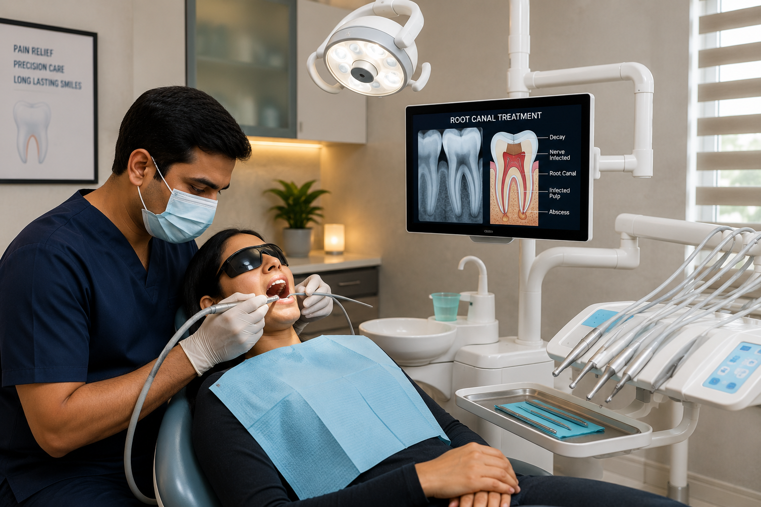
A digital RVG X-ray (or OPG for larger lesions) confirms the cyst's size, location and involvement with nearby structures.
Root canal treatment or retreatment removes the infected nerve tissue that is feeding the cyst — the essential first step in most cases.
Small cysts are monitored to heal after RCT. Larger or persistent cysts are treated surgically via apicoectomy or enucleation.
Surgically removed cyst tissue may be sent for biopsy to confirm the diagnosis and rule out other pathology.
Follow-up X-rays over the following months confirm that the bone around the root tip is healing and refilling normally.
Removing the infection source through root canal treatment often allows small cysts to heal on their own over time.
A minor surgical procedure to remove the infected root tip and cyst tissue when non-surgical treatment alone isn't enough.
Complete surgical removal of the cyst lining, typically used for medium to large cysts to prevent recurrence.
For very large cysts, a small opening is created to decompress the cyst gradually before final removal.
Cost depends on cyst size, complexity and whether surgery is needed. Full quote after X-ray review.
Root canal treatment or retreatment to eliminate the infection source.
Minor surgical removal of the cyst and infected root tip.
Lab analysis of removed tissue to confirm diagnosis.
Skilled at diagnosing and treating periapical pathology while prioritising saving your natural tooth.
Digital RVG X-rays accurately map cyst size and root anatomy for reliable treatment planning.
We always attempt non-surgical resolution first, reserving surgery for cases that truly need it.
Scheduled review X-rays track healing so you know your treatment has genuinely succeeded.
| Factor | Periapical Cyst | Dental Abscess | Granuloma |
|---|---|---|---|
| Nature | Fluid-filled sac | Acute pus collection | Chronic inflamed tissue |
| Pain Level | Often none | Severe | Mild or none |
| Onset | Slow, months–years | Rapid, days | Slow, chronic |
| Typical Treatment | RCT ± surgery | Drainage + RCT/antibiotics | RCT, monitored |
Cyst treatment is closely linked to Root Canal Treatment and Laser-Assisted RCT — explore these for related endodontic care.
Book a consultation at PRALASH Dermystique & Dentique, HRBR Layout — early treatment saves your tooth and your bone.