At PRALASH Dermystique & Dentique, HRBR Layout, digital RVG X-rays give you a clear, detailed image in seconds — with a fraction of the radiation of traditional film, for safer, faster diagnosis.
RVG stands for RadioVisioGraphy — a digital intraoral X-ray system where a small sensor placed in your mouth captures the image directly onto a computer screen, instead of using traditional film that needs to be chemically developed.
The result is a sharper, faster, lower-radiation X-ray that can be zoomed and enhanced on screen — used alongside our Intra-Oral Scanners and CBCT imaging to build a complete digital picture before treatment planning.
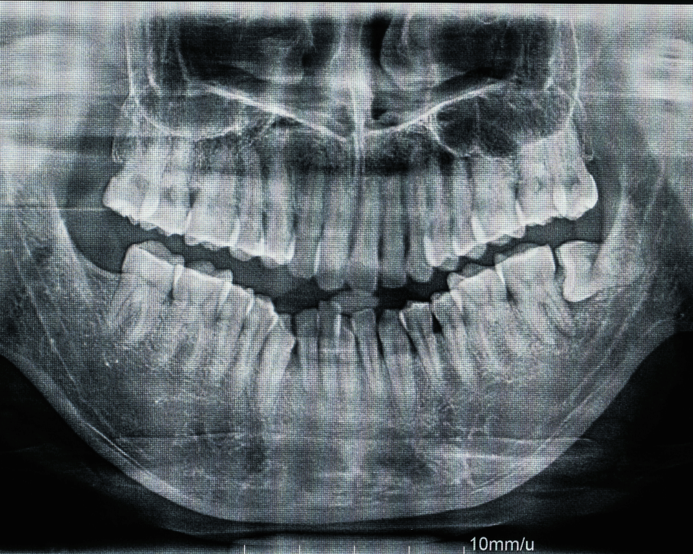
A small, flat digital sensor is placed against the tooth or area being examined.
A very short, low-dose exposure captures the image — over in a fraction of a second.
No developing, no waiting — the image is visible on the computer within seconds.
Brightness, contrast and zoom are adjusted on screen for the clearest possible diagnostic view.
Your dentist reviews the image with you in the same visit, pointing out exactly what it shows.
Images are stored digitally, making it easy to compare against future X-rays over time.
Shows a full tooth from crown to root tip — used to check root canal work, infections and abscesses.
Captures upper and lower teeth together — the standard way to check for cavities between teeth.
A complete set of images covering every tooth, used for comprehensive treatment planning.
A quick check X-ray to confirm a filling, crown, root canal or extraction site is healing as expected.
A fraction of the exposure of traditional film, for a diagnostically clear image.
No darkroom, no waiting — the image appears on screen within seconds.
Digital tools let your dentist magnify and adjust the image for a more precise diagnosis.
Digital records make it simple to track changes in a tooth or bone level over time.
A cleaner process for the clinic, with nothing to develop or dispose of.
An RVG X-ray is typically recommended for:
Cost depends on how many images are needed for your diagnosis or treatment.
One periapical or bitewing image for a specific tooth or area.
A complete set of images for comprehensive treatment planning.
Digital sensors are used clinic-wide, keeping every X-ray as low-dose as possible.
Images are reviewed with you immediately, so treatment decisions aren't delayed.
From routine checkups to root canals, implants and orthodontics — imaging is built into the process.
Easy to reach from HRBR Layout, Kalyan Nagar and Kammanahalli for a quick diagnostic visit.
| Factor | Digital RVG | Traditional Film |
|---|---|---|
| Radiation Dose | Up to 80–90% lower | Higher |
| Result Time | Seconds, on-screen | Minutes, needs developing |
| Image Adjustment | Zoom, enhance digitally | Fixed once developed |
| Storage | Digital patient record | Physical film, can degrade |
| Environmental Impact | No chemicals | Requires developing chemicals |
RVG X-rays are a routine part of Root Canal Treatment and Guided Implant Rehabilitation — see how imaging fits into each treatment.
Book a consultation at PRALASH Dermystique & Dentique, HRBR Layout — digital RVG imaging is part of every diagnostic visit here.